

Up to 10% of people in their later years can be affected by an epiretinal membrane (ERM). Here’s what to watch out for and how to manage it.
What is an Epiretinal Membrane?
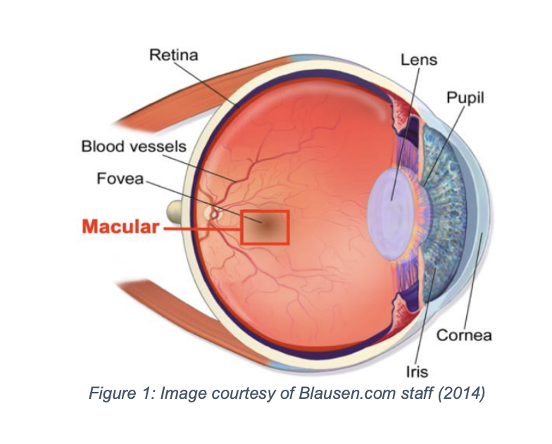
Epiretinal membrane (ERM) is a condition where a thin layer of scar tissue forms on the retina’s surface, leading to blurred and distorted vision. It affects the macula, the part of the eye responsible for sharp, central vision used in activities such as reading or driving. The development of an ERM can cause the macula to crumple or tighten, distorting your sight.
This diagram highlights the retina, where ERM forms, and the macula, where vision is affected.
Why does an ERM appear?
In most cases, an ERM develops due to natural ageing. However, they can also be triggered by diabetes, eye inflammation, blood vessel blockage, or previous eye surgeries. Some studies suggest that women have a higher risk. Typically, only one eye is affected.
Suspect you may have an ERM? Here’s what to do
Your optician can diagnose ERM during an examination of the retina. Your vision and eye pressure will be checked. It may be necessary to use eye drops to dilate your pupils for further examination. A scan of the back of your eyes may also be performed. Your symptoms will be assessed before deciding the next course of action.
Diagnosing an ERM
ERM is often discovered by chance, especially if it hasn’t yet affected vision. In such cases, it may never worsen. However, if it does cause significant visual distortion or blurriness, treatment options will be explored. It is important to know that even untreated, an ERM will not lead to complete blindness. Often, regular monitoring by your optician is sufficient.
Treatment
If treatment is needed to combat the growth of the membrane, then it can be treated with vitrectomy surgery.
Vitrectomy is a procedure where the vitreous jelly inside the eye is removed, and the ERM is peeled away from the back of the retina. The vitreous jelly is not replaced; however, a natural fluid will replace the vitreous jelly over time.
The operation typically takes less than an hour and can be done under local or general anaesthesia. Recovery can take several months, but 70-80% of patients experience vision improvement within 3-6 months. Most people take about two weeks off work post-surgery, with a follow-up appointment after the procedure.
If you think you may have an epiretinal membrane or have noticed changes in your vision, please call us to book an appointment.
Helpful resources
RNIB
Website https://www.rnib.org.uk/
Helpline – 0303 123 9999
The Macular Society
Website https://www.macularsociety.org/
Helpline – 0300 3030 111
Sources
Imperial College NHS
National Library of Medicine
Guide Dogs